Tracheal stenosis is the narrowing of the trachea due to the formation of scar tissue or malformation of the cartilage in the trachea. Mild narrowing of the trachea may not cause complaints. When the narrowing is more than 50 percent of the airway, serious complaints can cause serious complications.
The three most common causes of tracheal stenosis are:
- Prolonged insertion of an endotracheal tube (breathing tube) or tracheostomy
- Compression of tumors and masses adjacent to the trachea
- Goiter and Thyroid cancers
- Lung cancer
- Lymphoma
- Congenital malformations (congenital disability)
- Radiation therapy
- Tracheal infections
- Inhalation burns
- Inflammatory diseases (sarcoidosis or amyloidosis)
- Cancer
- Tracheal tumors
- Collagen vascular disease (granulomatosis with polyangiitis)
- Trauma
In cancer and congenital malformations, the airway is compressed by narrowing the trachea or malformed cartilage.
Other causes of tracheal stenosis usually begin with ulceration of the trachea. The ulcer results from inflammation. The additional scar tissue narrows the space in the trachea.
In patients who have been intubated for a long time, necrosis develops if the balloon of the intubation tube is too swollen and stays for a long time. Scar tissue occurs, and the tracheal lumen becomes narrowed.
Tracheal stenosis due to prolonged incubation in intensive care is around 1-2% and may occur after three months.

Figure 1: Trachea anatomy

Figure 2. Tracheal Stenosis in Thorax CT
Tracheal Stenosis Symptoms
In congenital tracheal stenosis, mild stenosis can present as asthma or recurrent bronchitis. In mild tracheal stenosis, the patient may not identify symptoms until later childhood or early adolescence. In more severe cases of congenital tracheal stenosis, we may see the following symptoms:
- Stridor (high-pitched breathing sound)
- Cyanosis, with prominent blue lips
- Wheezing
- Shortness of breath (dyspnea)
In other cases of acquired tracheal stenosis, symptoms appear several weeks after the injury. Difficulty breathing is the typical first symptom. Stridor, wheezing, or shortness of breath on exertion are common.
Diagnosing Tracheal Stenosis
- Various methods help us diagnose tracheal stenosis.
- Bronchoscopy is considered the "Gold Standard" for diagnosing tracheal stenosis because it directly visualizes the trachea.
However, there are some risks because a scope further obstructs your airway, affecting oxygenation levels. Remember to discuss your risk factors associated with bronchoscopy with your doctor.
Other methods include X-ray, Thoracic CT scan, ultrasound, MRI, and pulmonary function testing.
A CT scan determines tracheal stenosis. However, it is challenging to identify soft tissue diseases of the trachea. Some techniques are used to create a "virtual endoscopy" to minimize the need for bronchoscopy. However, CT scanning is not an excellent method for determining less severe stenosis.
Ultrasound can be helpful in determining the amount of air space in the trachea.
How Is Tracheal Stenosis Treated?
Tracheal stenosis, including subglottic stenosis, is a narrowing of the trachea that causes breathing problems. It forms scar tissue in the trachea due to prolonged intubation -- when a breathing tube is inserted into the trachea to maintain breathing during a medical procedure -- or from a tracheostomy, which is an operation to create an opening in the neck. The stenosis may require surgery. The type of procedure depends on the exact location and degree of stenosis.
The most common treatment options are:
Bronchoscopic Tracheal Dilatation: Enlargement of the trachea with surgical instruments called balloons or tracheal dilators provides temporary relief of symptoms and allows our specialists to determine the severity of stenosis. During the dilation procedure, we can diagnose the cause of the stenosis.
Laser Bronchoscopy: In some cases, surgeons use lasers to remove the scar tissue causing the stenosis. Laser surgery offers good short-term results and temporary relief but is not usually a long-term solution. In some cases, laser surgery can worsen the stenosis. For these reasons, it is essential to consider the underlying disorder before laser surgery.
Tracheobronchial Airway Stent: A tracheal stent is a tube made of metal or silicone inserted into the airway to keep it open. Stents are used as both short-term and long-term therapy for stenosis.
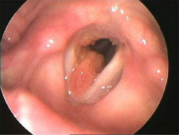
Figure 1. Tracheal silicone stent
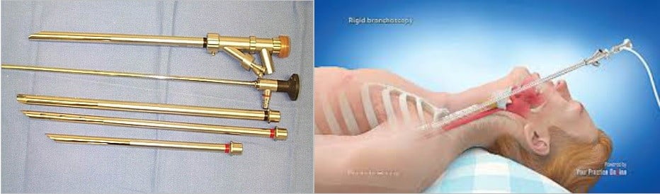
Figure 2. Rigid Bronchoscopy
Tracheal Resection and Reconstruction: During tracheal resection, our surgeons remove the narrowed section of the trachea and then reattach it to the upper and lower areas. It is a very successful treatment for stenosis with excellent long-term results.
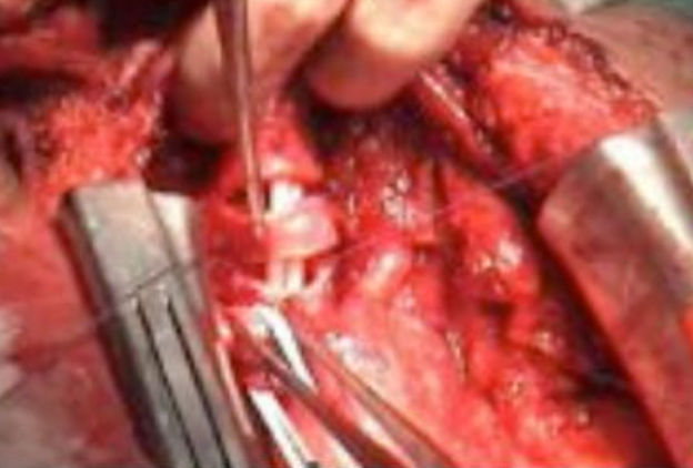
Figure 3. Tracheal segmental resection

Figure 4. Suturing the cut trachea to three planes
Tracheal Stenosis – Frequently Asked Questions About Tracheal Stent
Tracheal stenosis is the undulation of the windpipe that occurs after radiation therapy, prolonged use of the breathing tube, or other procedures. Show details. Tracheal stenosis, including subglottic stenosis, is tracheal undulation that causes breathing problems.
Symptoms of tracheal stenosis are typically: Wheezing, coughing or shortness of breath, including difficulty breathing. A high-pitched wheezing that comes from your lungs when inhaled.
Chest X-ray: A plain X-ray can tell us if the trachea has slipped on both sides of the chest. Chest radiography is not enough. Thorax CT required. It is necessary to request Thorax CT (Picture 2) with reconstruction for the retracted trachea. If it is not said so, you may have to take it again.